Friday, March 31, 2017

Altered mental status, seizure, extreme hypertension, and a bizarre ECG

A middle aged woman with few serious medical issues presented with altered mental status, had a generalized tonic clonic seizure, and was found to be hypertensive and tachycardic.  An ECG was recorded:
What is it?
The computer measures QRS duration as 126 ms.


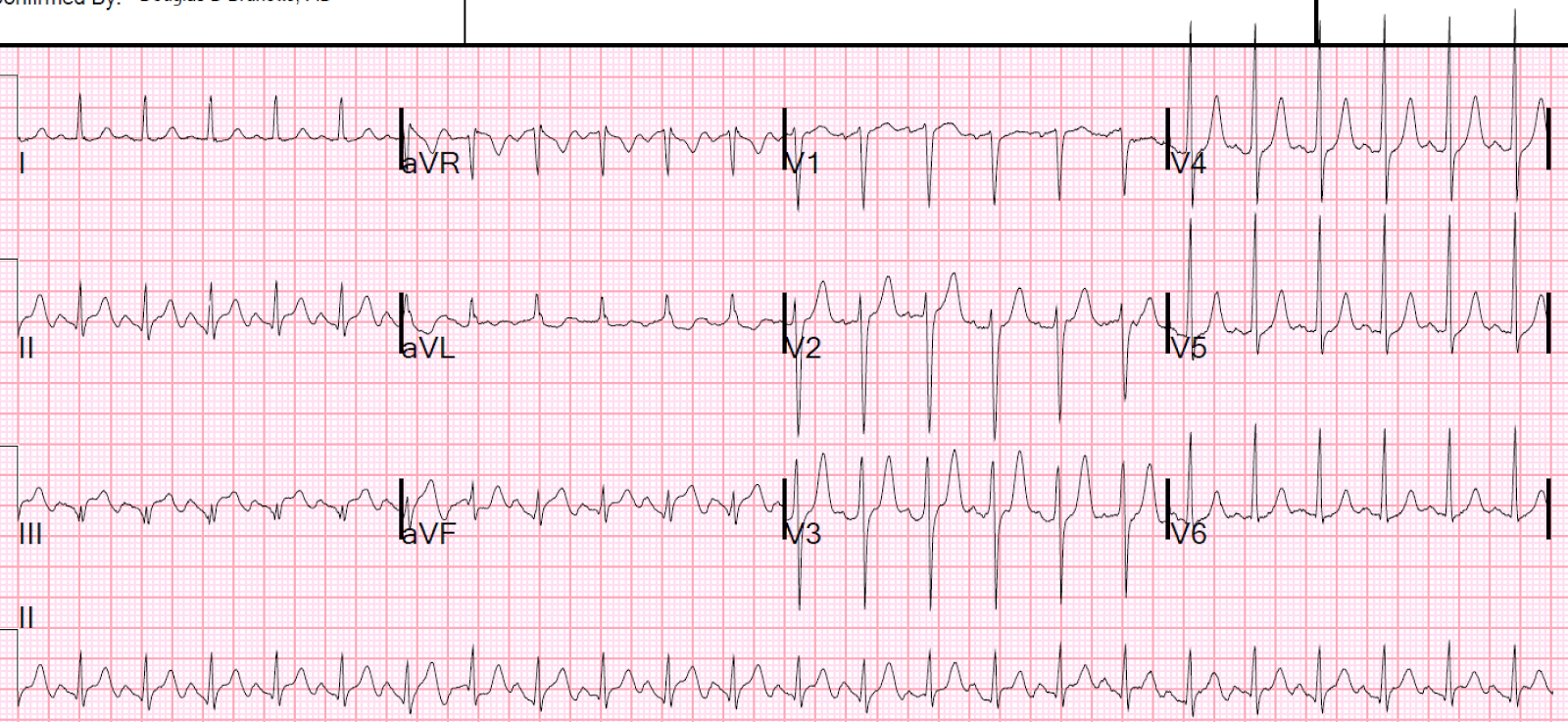
This is a regular, wide complex tachycardia, but not very wide.  There are no P-waves.  The QRS has 2 alternating morphologies, both of which are right bundle branch block (RBBB) in configuration, but which have different axes.  Both complexes have an rSR' in lead V1 and a wide S-wave in V6.

One might suspect that the higher voltage beats are premature beats, but by my calipers, the intervals are identical.  (This is critical to being certain that they are not premature beats.)  Similarly, one might suspect every-other-beat pre-excitation, but no delta waves are seen.

See how the S-wave in lead I alternates small, large, small, large...  The large S-wave indicates right axis deviation and appears to be due to alternating left posterior fascicular block, in addition to RBBB.

So this is SVT with alternating aberrancy: RBBB, then RBBB + LPFB, then RBBB, then RBBB + LPFB.....

The question of electrical alternans came up.  SVT frequently has electrical alternans but, unlike in sinus tach, it is not correlated with effusion or tamponade.  In this case, it is not the typical SVT electrical alternans -- the alternans is due to alternating conduction.

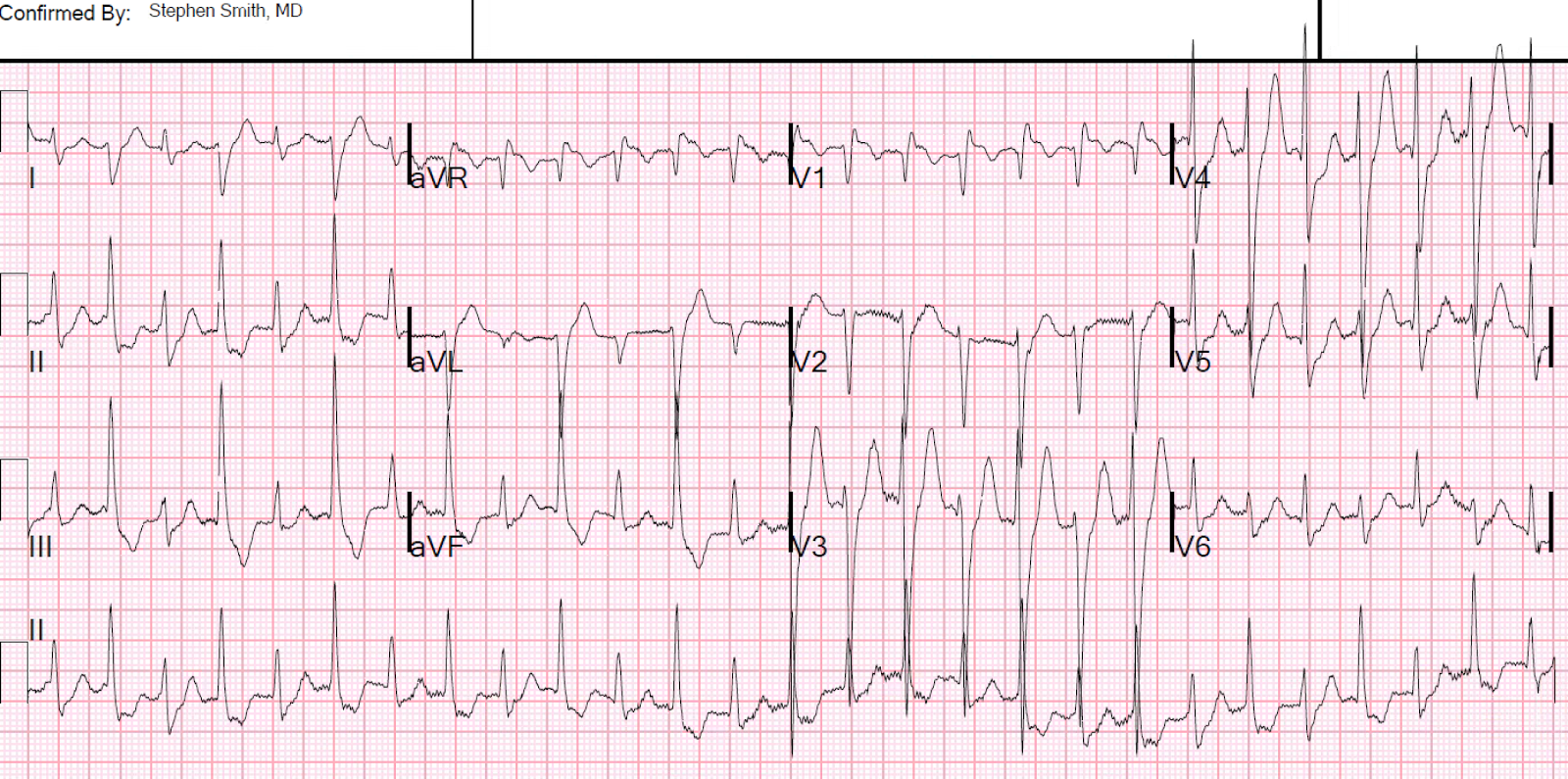
She spontaneously converted:
Sinus tachycardia with normal conduction.
Normal QRS, narrow, normal axis.
This confirms that the SVT caused the aberrancy.

ST depression consistent with ischemia.


She was found to have a BP of 300/180, and sinus tachycardia at a rate as high as 150.  This blood pressure and heart rate were successfully controlled with clevidipine and esmolol.  The patient was found on MRI to have "Posterior Reversible Encephalopathy Syndrome" (PRES), which is not always posterior and not always reversible, and which formerly was called "hypertensive encephalopathy."  The extreme hypertension and sinus tachycardia led to suspicion of pheochromocytoma.

The etiology of the SVT is uncertain, but is probably AVNRT that was initiated in this hyperadrenergic state.  The aberrancy is due to refractoriness of the right bundle at this high rate, and a posterior fascicle which is only able to recover on every other beat, but is still refractory on the beats inbetween.  The right bundle nearly always has a longer refractory period than the left bundle and aberrancy is thus much more likely to show RBBB morphology.


Here is another case of SVT with alternating aberrancy


An Unusual Tachycardia


Is there an alternative explanation?  

Bidirectional Ventricular Tachycardia may be due to a "ping-pong" effect of alternating VT origin in the left bundle vs. right bundle.  It is conceivable that this could be fascicular VT with an alternating origin in the left bundle, then left anterior fascicle, back and forth.  Then it would be a bidirectional fascicular VT.  I've never heard of this before and could not find any literature on it, so it probably doesn't exist and this hypothesis is pure speculation.  See Ken Grauer's comments on this differential diagnosis, pasted below.  As always, it is very incisive!

Ken Grauer's comments

I'd bet this is alternating RBBB with RBBB/LPHB aberration. I had not heard of bidirectional fascicular VT — and I'm sure it is rare indeed. My impression from the fascicular VTs that I've seen — is that although they resemble some pattern of bifascicular block (rbbb/lahb or rbbb/lphb) — they are usually NOT as "clean" as can be the case with aberrancy or preexisting bbb. 

Of course, aberrancy and preexisting bbb/hemiblock patterns are not always "clean" (they may show squiggle, notches, lack that clear S wave in V1, etc) — but when you do see a bbb or bifascicular block pattern that is "clean" — my impression is that it is almost certain to be supraventricular; and not ventricular in etiology. 

I believe that is the case here. In some leads (like lead II) — you'll note not only the initial direction, but also the initial slope of the initial deflection is identical for both complexes. If we were switch from one site of fascicular VT to another — I would not expect to see this. In other leads — the very fine detail of the initial deflection is doing exactly as I would expect it to do if the problem was alternating LPHB with every other beat. That is, in lead I with LPHB beats — there is a tiny-but-present initial r wave — but instead there is a tiny-but-present initial q wave when we only have RBBB. And in lead III with LPHB beats, there is a tiny-but-present initial q wave — but with pure RBBB beats in lead III there is no such initial q wave. This is precisely what is supposed to happen when you have isolated LPHB. Similarly, in lead V1, there is a tiny-but-present initial r wave for pure RBBB beats — but this initial tiny r wave is lost with LPHB beats in V1. And then in V2,V3 the R wave is decidedly taller with the LPHB beats — which is as expected (the only functioning fascicle is the LAH, so the QRS is more positive in V2,V3 reflection more anterior depolarization that is no longer opposed by the block LPH).


Ken

Tuesday, March 28, 2017

Echocardiography, even (or especially) with Speckle Tracking, can get you in trouble. The ECG told the story.

A completely healthy 30-something year old woman with no cardiac risk factors had sudden onset of bilateral trapezius pain that radiated around to her throat.  It resolved after about 5 minutes, but then recurred and was sustained for over an hour.  She called 911.

EMS recorded these ECGs:

Time 0:
In V2-V4, there is ST elevation that does not meet STEMI "criteria," of 1.5 mm at the J-point, relative to the PQ junction.  But there are also unusually Large T-waves

Called Normal by computer
This is the Zoll algorithm


I sent this to the Queen of Hearts:


Time = 13 min
T-wave in V2 is now taller and fatter, the ST segment is more straight.
T-wave in V3 is no taller, but it is fatter due to a straighter ST segment
This is highly suspicious for early LAD occlusion

Not identified as STEMI, but not normal either
This is the Zoll algorithm


Time = 24 min
No significant change
Zoll did not recognize acute MI, but did not call it normal

These prehospital ECGs were not seen.

The patient arrived in the ED.

The pain completely resolved after nitroglycerine 


Moments later, the this ECG was recorded in the ED when she had been pain free for moments only:
Computer read: Normal ECG.

This was by the Veritas algorithm

The ECG is diagnostic of LAD OMI

However, T-waves are still unusually large; the computer almost never sees this.
The T-wave in V2 is smaller. 
QTc is 444 ms.
STE 60 V3 = 1.5 mm, R-wave amplitude V4 = 15 mm, QRSV2 = 8.5
Formula value is 19.38, which indicates LAD occlusion 
(The most accurate cutpoint is 18.2 -- a value greater than 18.2 has high probability of LAD Occlusion).  

This patient has a nondiagnostic ECG by most rules.  

However, with attention to subtleties, especially when compared with the unseen prehospital ECGs, it is very worrisome.

And to my expert pattern recognition it is definitely LAD OMI


Since I taught the Queen of Hearts, she knows that it is OMI:


The Queen of Hearts PM Cardio App is now available in the European Union (CE approved) the App Store and on Google Play.  

For Americans, you need to wait for the FDA.  But in the meantime:

YOU HAVE THE OPPORTUNITY TO GET EARLY ACCESS TO THE PM Cardio AI BOT!!  (THE PM CARDIO OMI AI APP)

If you want this bot to help you make the early diagnosis of OMI and save your patient and his/her myocardium, you can sign up to get an early beta version of the bot here.  It is not yet available, but this is your way to get on the list.


The first troponin was below the level of detection (LoD).

If you use something like the HEART score:
1. H  History: She has atypical pain (trapezius) (score = 0)
2. E  EKG: a negative ECG (score = 0)
3. A  Age: = 0
4. R  Risk factors = 0
5. T:  Troponin = 0 [first troponin (contemporary, not high sensitivity) was less than the level of detection). 
Total HEART score = 0.  Risk of 30-day adverse events is less than 1.7%.   Some might send her home.

But maybe she has an acute LAD occlusion that will get even worse. 


The providers did a bedside echo and even used speckle tracking to look for strain:

I think maybe there is an anterior wall motion abnormality, but this is very difficult.  They read it as normal.

Here are a couple shots with strain, or "speckle tracking" on ED Echo:


To, me these look like anterior wall motion abnormality, but I showed them to one of our ultrasound fellows who is very interested in this.

She said:

This is a tough one. I see what you mean, initially when I looked at the image, I also thought there was an anterior wall motion abnormality.  But then on closer inspection, I suspect that maybe the anterior wall is just not being tracked well. In systole, you can see the anterior wall come down and outside of the area that is being tracked (more so than the other tracked walls). Even though the strain values are a little off in the graph (so is the posterior wall) it is still a value range (about -18) that would be considered non-ischemic by the cardiology literature, I believe.  I have been wrong before though! So it is possible that I am misinterpreting the clip. If it were me, I would get values at the level of the mitral valve, papillary muscles, and apex (all in PSS axis). Also, narrowing the area being tracked helps the walls get recognized much better.


As I wrote, the first troponin was below the Level of Detection.

She remained pain free, and was admitted without further serial ECGs.  

When in doubt, one should always get serial ECGs.  Bedside echo is not enough.


At time = 240 minutes (4 hours), the second troponin returned at 1.15 ng/mL.  That prompted recording of this ECG:
Back to normal for this patient.  This demonstrates that all ST elevation of the previous ECGs was ischemic, not normal.  She was having a transient STEMI, briefly.

It is very lucky that she spontaneously reperfused her LAD.  It did not progress to full STEMI with loss of the anterior wall, as in this case.

Also, persistence of a pain free state does not guarantee an open artery.  See this case.

A formal contrast echo was done at this point:
Normal estimated left ventricular ejection fraction, 65%.
Regional wall motion abnormality-distal septum and apex.

She was treated medically for NonSTEMI, pending next day cath, which showed  ulcerated plaque and a 60% thrombotic stenosis in the LAD distal to the first diagonal.  It was stented.


Learning Points:
1. Always get serial ECGs when there is any doubt about what is going on.
2. Use the 4-variable formula!!

12 Example Cases of Use of 3- and 4-variable formulas, plus Simplified Formula, to differentiate normal STE from subtle LAD occlusion

3. Always find and look at prehospital ECGs.  They give extremely valuable information.
4. Hyperacute T-waves remain for some time after reperfusion of an artery.  I always say that "you get hyperacute T-waves both 'on the way up' (before ST segment elevation) and 'on the way down' (as ST elevation is resolving).
5. Wall motion abnormalities are very hard to see, even with advanced Speckle Tracking technology.  They require a great contrast exam and expert interpretation.
6.  This case does not demonstrate it, but a wall motion abnormality may disappear after spontaneous reperfusion (see this case).
7. Patients with transient occlusion may manifest only transient STEMI on ECG.  Subsequent troponins may be all negative and subsequent formal echo may be normal.  See this case


Friday, March 24, 2017

A young man with sudden chest pain

A 30-something with history of 2 pack per day smoking complained of sudden left arm and chest pain while working construction.  It was very distressing for him.  He presented by private transportation, stating that his pain was decreasing.

At triage, he had this ECG recorded:
The computer read that there is incomplete right bundle branch block.
QRS duration 102 ms.
What do you think?


No significant abnormalities were seen in triage, and the patient had to wait 2 hours.  By the time he was roomed, his pain was gone.

A second ECG was recorded, pain free:
If you didn't see anything wrong with the first one, maybe you can see it now by comparison with this one.


His first troponin I, drawn 4 hours after arrival, was 1.6 ng/mL.


Analysis:
--There is subtle ST Elevation in inferior leads II, III, aVF, with reciprocal ST depression in aVL.  This is diagnostic of inferior injury.  The fact that it resolved with resolution of chest pain simply verifies this.
--There is also a decrease in the size of the lateral precordial T-waves.
--Notice that the ST elevation on the first ECG does NOT meet STEMI criteria. There is not 1 mm of STE in any lead.
--But that ECG is diagnostic of injury.


The patient was started on heparin.

Shortly thereafter, he had a run of ventricular tachycardia.  Therefore he was taken urgently to the cath lab.  [Patients with ACS who have persistent refractory pain, hemodynamic or electrical instability, or pulmonary edema, should go emergently to the cath lab]


The angiogram was clean!!  

An MRI was diagnostic of myocarditis.  

--There were no wall motion abnormalities (although they are frequently seen in myocarditis)
--No evidence of microvascular obstruction
--Delayed enhancement sequences obtained at 10 mins after gadolinium administration reveal multiple (at least 3) foci of delayed enhancement, measuring about 2 centimeters in the 
inferior septum, and approximately the same size in the inferior wall.
--There is subendocardial sparing demonstrated (this pretty much establishes myocarditis, as ischemia would preferentially affect the subendocardium.

Troponin I profile:

Time after arrival         Troponin I
4                                     1.613
6.5                                  5.887
13.5                              10.969
15.5                              11.347
19                                   9.001
21.5                                7.521
25                                   6.175  


Learning Points:

1.  Learn to recognize subtle injury. This could have been an early STEMI.  The patient's reperfusion time would have been delayed.
2.  It is particularly easy to miss these when you are shown an ECG completely out of clinical context, as often happens with triage ECGs.
3.  Injury NOT due to ischemia also is high risk: in this case, the risk of ventricular dysrhythmias.
4.  Myocarditis and STEMI are often indistinguishable.  They may have injury on ECG, wall motion abnormalities, and elevated troponins.  ACS may also have a negative angiogram if there is autolysis of thrombus!  MRI makes the diagnosis.

Here is another interesting related case:

A Young Woman with Chest Pressure and Subtle, Focal ST Elevation/Depression

The effect of targeted temperature management on QT and corrected QT intervals in patients with cardiac arrest


Just published online:


Tuesday, March 21, 2017

2 different wide complex rhythms (rate about 60) in a patient with chest pain

A male in his 40s presented to the ED by ambulance with acute chest pain, possibly waxing and waning.  He had been working out and felt dizzy and weak in addition to the chest pain.

He had a pacemaker in place that he stated was due to a congenital problem, later found to be due to congenital absence of atrial activity.  He had no history of coronary disease.

Here is his first ED ECG (ECG 1):
What do you think? (It is NOT paced)
See analysis below.


ECG 2: 25 minutes later
What do you think? (Still not paced)
See analysis below.


ECG 3: His prehospital ECG was located; it had been recorded 25 minutes before the first ED ECG:
What do you think?


ECG 1: 

1. There are no P-waves.  The rate is slightly above 60.  There are no pacer spikes, so either the pacer is not working or its rate is set below 60.
2. There is an RBBB and LAFB morphology, which means that either the junction is escaping and there is RBBB + LAFB, or there is a posterior fascicular escape.  A rate of 60 suggests that it is junctional and not fascicular.
3. V2 lacks the normal ST depression that one sees in RBBB.  In RBBB, the ST segment is usually discordant (opposite to) the positive R'-wave, with up to 1 mm of ST depression, also usually with an inverted T-wave, as seen in the "normal RBBB" ECG below.
4. Concordant STE in lead I
5.  Notice the slight ST elevation in V3, with up-down T-wave. This is highly suggestive of ischemia.
6.  There is very poor R-wave progression from V3-V6.
7.  There are Proportionally large T-waves from V3-V6.

In this situation, all these features are all but diagnostic of LAD occlusion.


Normal RBBB
Normal RBBB, with normal ST depression, up to 1 mm, in V2 and V3, and inverted T-waves.
Note the absence of ST elevation and the size of the T-waves in lateral precordial leads.


ECG 2:

Same analysis, except that now there is new ST elevation in V4-V6 and dynamic T-waves in V2 and V3, with slightly increased ST elevation in aVL with reciprocal ST depression in lead III.


ECG 3: Prehospital ECG:

1. This one is paced!! (Also at a rate slightly above 60)
2. Look at lead V6.  There is proporionally excessively discordant ST elevation at 2.5 mm.  Divide that by the preceding S-wave (6 mm), and the proportion of 2.5/6 = 42%.  I believe this is diagnostic of coronary occlusion.
3. The T-waves in V3-V6 are very abnormally large, highly suggestive of acute occlusion.

ECG in Ventricular Paced Rhythm with Coronary Occlusion

The Modified Sgarbossa criteria for left bundle branch block (LBBB), which I apply to ventricular paced rhythm, uses a hard cutoff of 25% in just one lead to diagnose coronary occlusion.  This is the third criterion (1st criterion is concordant STE of 1 mm in 1 lead, and 2nd is concordant ST depression of 1 mm in one of leads V1-V3); any one of the 3 makes the diagnosis.  Our research would indicated that the normal discordant proportional ST elevation is about 11% and that anything above 15% is very likely to be occlusion.  A 25% ratio was 99% specific, but a 20% proportion was still 94% specific in the validation study!

We are in the midst of a multicenter, international study to assess the Modified Sgarbossa Criteria in Paced Rhythm.  It is called the PERFECT study!  (Paced ECG Requiring Fast Emergent Coronary Therapy).  https://clinicaltrials.gov/ct2/show/NCT02765477


Many physicians still do not believe that STEMI can be diagnosed in the presence of ventricular paced rhythm.

One of my mentors, K. Wang, put up an image on Medscape of a Paced ECG that clearly showed STEMI and quizzed the readers and received this answer:
50% gave the wrong answer: you cannot diagnose infarction in ventricularly paced rhythms.


Case Continued:

The cath lab was activated emergently and the patient was found to have 100% occlusion of the first Diagonal off the LAD.  It was opened and stented.

The peak trop I was 33.9 ng/mL.

A subsequent echo showed:


--Normal left ventricular size, thickness estimated left ventricular ejection fraction is 50-55 %.
--Regional wall motion abnormality-distal septum anterior and apex,dyskinetic.
--Regional wall motion abnormality-distal inferior wall, dyskinetic.
--Asynchronous interventricular septal motion nonspecific.


Learning Points:
1. RBBB can obscure LAD occlusion, but you should suspect it in the absence of normal discordant ST depression in V2 and V3, or with hyperacute T-waves.
2. Paced rhythm obscures MI far less than believed.  Coronary occlusion can definitely be diagnosed in the presence of ventricular paced rhythm.
3. Even in normal conduction, acute coronary occlusion is frequently not evident on the ECG. This is also true with RBBB, LBBB, and Paced Rhythm.  There is little evidence supporting the notion that it is far more difficult in the latter three.

Sunday, March 19, 2017

Tachycardia, Dehydration, and New ST Elevation in a 20-something, then a Surprise.


A male in his early 20s presented intoxicated, with no CP or SOB.  An ECG was obtained for tachycardia.

First ECG:
Sinus tachycardia at 150.
There is inferior ST Elevation and Q-waves that appear to be Inferior STEMI.
There is reciprocal ST depression in aVL.
Is this STEMI?


The patient was very dehydrated.

The physicians were worried about STEMI, and so did a Point of Care Cardiac Ultrasound which showed IVC collapse and hyperdynamic function.

The patient had a previous visit with dehydration
There had been some, but less, STE
Not very similar to this one.


There was another previous visit with dehydration
Also not similar


He was given 2 liters of IV fluids.  A 2nd ECG was recorded 1 hour later:
Sinus tach at a rate of 120
Heart rate and ST segments are almost normalized 


What do you see now?
See below.


Unexpectedly, there is every other beat pre-excitation.  So the patient has WPW. This was never diagnosed before.  But I don't think this explains his ST segments.

Clinical course

3 serial troponins were below the 99% level of 0.030 ng/mL, but not undetectable.

K was 4.5 mEq/L.

The patient was rehydrated and discharged.

Comment:

Why the PseudoSTEMI pattern?

I don't know for certain, and this ECG sure looks like inferior STEMI.  However, patients with STEMI generally do not have tachycardia unless they are in cardiogenic shock.  If such is the case, the patient should have high filling pressures and high right sided pressures and NOT have a collapsed IVC.  They should also not be hyperdynamic.

It is possible to be dehydrated AND have a STEMI, but in that case the best initial treatment is supportive: rehydration.  Then re-evaluation.

So it was perfectly appropriate to do a bedside ultrasound and, finding a hyperdynamic heart, to defer diagnosis of ACS and give fluids.  Even if this STE is due to ischemia, it is most likely due to demand ischemia, NOT due to ACS, and the best initial therapy is to hydrate.  In our many studies of type 2 MI, we found that 2-5% had ST elevation on the ECG.

Final comment:

I sent this to Ken Grauer, who is a fantastic ECG resource and has much experience with stress ECGs, and asked if this is a pattern he has seen in stress tests.  He stated "no," (in a few more words than that).

Friday, March 17, 2017

A 20-something with DKA and a regular wide complex tachycardia

Medics were called to see a 20-something type 1 diabetic with h/o DKA who had altered mental status.

He was found to have "Kussmaul" respirations and respiratory distress.

He was not in shock, his blood pressure was adequate, and pulses were strong.

He was put on the cardiac monitor:
Regular wide complex tachycardia.

A 12-lead ECG was recorded:
Regular Wide complex tachycardia at a rate of 200.
What do you think?
If you're the medic, what would you do?


Whenever there is a wide complex, especially in a patient with DKA, one should think of hyperkalemia (sinus rhythm, often with "invisible P-waves").

However, this rate is too fast for sinus tach and the morphology is not right for hyperkalemia.  Still, calcium is harmless and it is harmless to give 3 grams of calcium gluconate.

He arrived in the ED in the same condition.  He was hemodynamically stable but with altered mental status and respiratory distress.  Cardiac echo showed hyperdynamic performance.   Blood gas showed severe acidosis, with pH 7.00, pCO2 25 and HCO3 of 6.   K retured at 4.5 mEq/L.

He was treated with calcium, bicarbonate, and magnesium.  He was intubated.

He had this ECG recorded:
What do you think?
What do you want to do?
Suppose the patient did not have altered mental status from DKA and was not intubated?


Analysis: VT vs. SVT with aberrancy vs. sinus tachycardia with hidden P-waves.  (With a rate this fast, and no inkling of P-waves, sinus is very unlikely but if you were not sure, you could record Lewis Leads.)

This ECG is almost certainly supraventricular tachycardia with aberrancy.  Why?

First: pretest probability
The patient is young and without heart disease
Good LV function makes VT less likely (except for fascicular VT)

Second: the ECG itself:
1. There is an inferior axis.  The impulse originates cranially and propagates caudally.  In contrast, VT, because it originates in the ventricle, which is lower left, generally propagates toward the upper right.
2. More importantly, there is a typical right bundle branch block (RBBB) morphology:
    a. There is RR' in V1 and the R' is larger than the R.
    b. There is a rapid, narrow R-wave in V5 and V6, with a wide S-wave.

This latter makes SVT with RBBB aberrancy almost certain.

Thus, it is very likely that it will convert with adenosine.  The medics could have given adenosine and converted.  Since the patient was hemodynamically stable, there was no rush to convert and therefore it is OK that they left it alone.

If you're wrong and give adenosine to a patient with VT, don't worry.  Adenosine has been shown to be safe in Ventricular Tachycardia and is even recommended now in ACLS.

Of course, electrical cardioversion will work very well but generally requires sedation unless the patient is already obtunded.  This patient was intubated and sedated, so electricity is easy to use.

However, if this patient were not so sick with DKA that he needed to be intubated, adenosine would be the treatment of choice.

The patient was diagnosed with VT and electrically cardioverted.

Here is the post-cardioversion ECG:
Sinus Tachycardia with normal QRS and axis and nonspecific ST-T abnormalities


There is no baseline RBBB.  The right bundle generally has a longer refractory period than the left bundle, so that when there is a fast rate, it can result in RBBB aberrancy.  

The patient did well and was later diagnosed with SVT with aberrancy.


Learning Points:

1.  Adenosine is safe in regular wide complex tachycardia.  (But NOT in irregular polymorphic wide complex tachycardia, as this could be WPW with atrial fib)

2. If there is typical RBBB morphology, it is almost certainly SVT with aberrancy.  There are also fairly rare fascicular ventricular tachycardias which can mimic SVT with aberrancy.  However, you won't hurt them with adenosine.

Tuesday, March 14, 2017

Why did this patient have ventricular fibrillation?

A previously healthy 50-something collapsed while exercising.  He underwent CPR and defibrillation and was brought to the ED.

Here is his initial ED ECG:
What do you think?


Analysis
1. Sinus tachycardia due to post arrest state.
2. I see no evidence of STEMI or even of subtle coronary occlusion.  I see no evidence of ischemia.
3. In some leads, there appears to be a very long QT, but it lead II across the bottom is used, it becomes apparent that the P-wave is mimicking the T-wave in some leads.  QT is normal.
4. There is no evidence of structural heart disease or congenital ion channel disease.
5. There are non-diagnostic Q-waves in inferior leads.  These could represent old inferior MI, which provides the substrate (scar) for ventricular fibrillation.


The patient went for emergent angiography.  Here are the findings:

LAD with obstruction, TIMI-1 flow, opened and stented.  Other diffuse non-obstructive disease.


The peak troponin I was only 0.85 ng/mL.

The next day formal echocardiogram was totally normal (no old MI).


In November 2016, this study was published in Resuscitation:

Millin MG et al.  Patients without ST elevation after return of spontaneous circulation may benefit from emergent percutaneous intervention: A systematic review and meta-analysis.  Resuscitation 2016, Vol.108, p.54-60.


The authors looked at many studies of patients with out-of-hospital cardiac arrest (OHCA) in which angiogram findings were correlated with ECG findings.  They found that 1/3 of patients who did not have STEMI on the ECG had culprit lesions that "required intervention."  They caution that this does NOT mean that all patients with cardiac arrest without ST elevation need emergent cath lab activation.  They caution that it is not clear which of these patients do need the cath lab or how to identify which will have culprits that need intervention.  Further study is needed.

However, here is a subgroup of OHCA that definitely should go:

Previously healthy patients who have ventricular fibrillation without a clear cause.  This includes, but is not limited to, patients with ECG findings such as:
1) Old MI with scar (Q-waves diagnostic of old MI)
2) Brugada syndrome
3) Hypertrophic cardiomyopathy.
4) Evidence of arrhythmogenic RV dysplasia
5) Evidence of chronic cardiomyopathy (along with echo evidence of very poor LV function).  Most of these patients are not previously healthy.
6) Other?

If no definite etiology can be found, the ventricular fibrillation was likely due to coronary ischemia (from acute coronary syndrome, with plaque rupture and thrombus) that is not evident on the ECG.

Common coronary findings in patients with ACS that results in OHCA:

1. Fixed tight lesion(s) which, during exercise, resulted in ischemia (not ACS, intervention not always needed, at least not emergently)
2. Thrombosis which was occlusive or nearly occlusive, but is no longer so because of some spontaneous thrombus lysis (requires intervention).   
3. Culprit lesion which showered downstream platelet-fibrin aggregates, resulting in small vessel ischemia.  These culprits are dangerous because the thrombus can propagate again and occlude the artery (requires intervention).
4. Thrombus with persistently limited flow, as in this case, even thought the ECG that does not show evidence of ischemia (requires intervention).
5. Other?

Learning point:

ST Elevation, and especially ST elevation criteria, are not sensitive for ACS and not even very sensitive for coronary occlusion.  ST depression may also not be present.  A high pretest probability, such as unexplained ventricular fibrillation, is ample reason to activate the cath lab.


Friday, March 10, 2017

How unreliable are computer algorithms in the Diagnosis of STEMI?


A 61 year-old with chest pain arrived to the ED by ambulance with resolving chest pain.  Here is his ED ECG:
The computer interpretation was "normal"
What do you think?


This ECG is NOT normal.  The T-waves in V2-V4 are very large in proportion to the QRS.   They are suspicious for hyperacute T-waves. There is low QRS amplitude.  

Can you trust a computer interpretation of "normal?" 
---Most of the time, but obviously not all the time.


Years later, I sent this to the Queen of Hearts:

The Queen can see Hyperacute T-waves incredibly well.

The Queen of Hearts PM Cardio App is now available in the European Union (CE approved) the App Store and on Google Play.  For Americans, you need to wait for the FDA.  But in the meantime:

YOU HAVE THE OPPORTUNITY TO GET EARLY ACCESS TO THE PM Cardio AI BOT!!  (THE PM CARDIO OMI AI APP)

If you want this bot to help you make the early diagnosis of OMI and save your patient and his/her myocardium, you can sign up to get an early beta version of the bot here.  It is not yet available, but this is your way to get on the list.


  1. Hughes KE et al. Safety of Computer Interpretation of Normal Triage Electrocardiograms. Acad Emerg Med 2017; 24(1): 120 – 24. PMID: 27519772
This recent study, discussed on Salim Rezaie's fine site REBEL EM, implies you can trust the computer interpretation of "normal." (http://rebelem.com/triage-ecgs-reducing-interruptions-busy-ed/)

What to do?

Recording serial ECGs would be useful.  The chest pain is resolving, so if these are resolving hyperacute T-waves, then followup ECGs should show their size diminishing.

However, when I saw this patient, I knew that he had come by ambulance, so I knew there must be a prehospital ECG recorded somewhere and went to look for it.

I found it.

Here it is:
Need I say more?
Yes, I'll say more: see the computer interpretation:
"Normal variant ST elevation, consider early (repolarization)"
Why the medics did not see it, I'm not sure. 


The cath lab was activated, as it should be with transient STEMI.

See this case of transient STEMI:

Spontaneous Reperfusion and Re-occlusion - My Bad Thinking Contributes to a Death.


Angiogram: There was 80% obstruction of the LAD with a large thrombus and TIMI-2 flow; the thrombus was suctioned out.

All serial troponins were undetectable!

Comment:
Had we not seen those subtle hyperacute T-waves, and then sought out the prehospital ECG, the patient would have ruled out, with one of three outcomes:

1. Subsequent thrombus propagation with re-occlusion, recurrent chest pain and diagnosis made (although perhaps late and perhaps only after an adverse outcome)

2. Undergone stress test with uncertain results.

3. Been discharged to home with potential disastrous outcome.


Learning Points

1.  Computer algorithms that make the diagnosis of "normal" are usually correct, but is usually good enough?

2. Computer algorithms are completely unreliable at diagnosing STEMI, with both poor sensitivity and poor specificity.  Here are two recent articles confirming this:

    a. The Comparison of Physician to Computer Interpreted Electrocardiograms on ST-elevation Myocardial Infarction Door-to-balloon Times.
    b. Electrocardiographic diagnosis of ST segment elevation myocardial infarction: An evaluation of three automated interpretation algorithms

3.  Always look for the prehospital ECG

4.  Hyperacute T-waves occur not only shortly after onset of chest pain (as the ST segment is about to rise, or "on the way up"), but also shortly after reperfusion (as the ST segment is resolving after reperfusion or "on the way down").

5.  With very brief occlusions, troponins may all be negative.  What would high sensitivity troponins have shown?  We don't know.

Recommended Resources